Our directions

Research

Our research lies at the intersection of signal processing, deep learning, and medical imaging hardware. We pursue end-to-end innovation: from imaging physics and algorithms to real-time deployment on portable, wearable, and robotic devices.

01

AI for Medical Imaging

Analysis, measurement, and reliable deployment

Segmentation/detection-based automation of clinically relevant parameter measurement, and robust, trustworthy medical AI with calibration and uncertainty, controllability, and generalization for real clinical workflows.

Measurement automationSegmentation & detectionUncertainty & calibrationTrustworthy AI
H-BiSeNet-Q segmentation on the DPU and keypoint measurement on the APU of a single SoC → automatic fetal AC/HC/BPD/FL measurements (green) closely matching manual ones (red) · Expert Systems with Applications, 2024 (JCR top 7%)
H-BiSeNet-Q segmentation on the DPU and keypoint measurement on the APU of a single SoC → automatic fetal AC/HC/BPD/FL measurements (green) closely matching manual ones (red) · Expert Systems with Applications, 2024 (JCR top 7%) Paper ↗
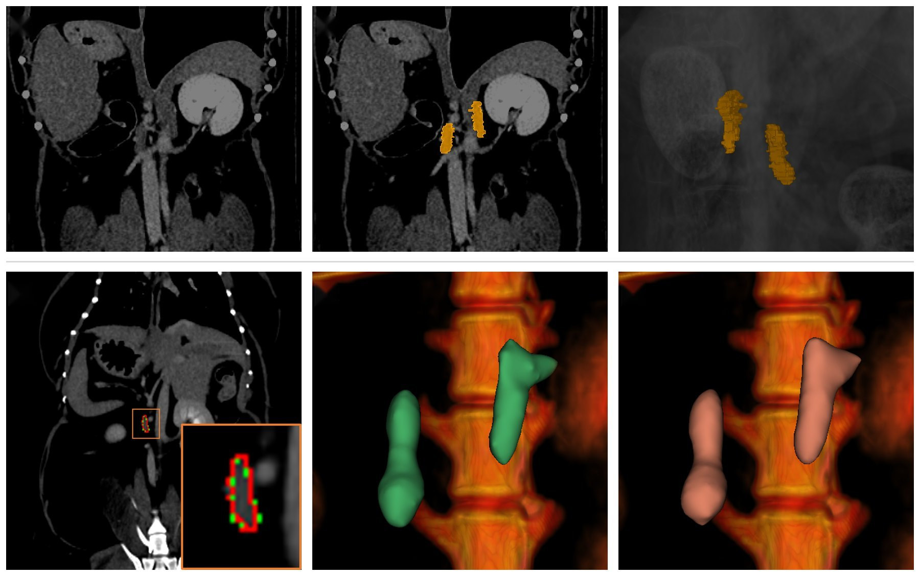
Manually annotated adrenal glands (top) train a Swin-UNETR 3D segmentation model → prediction (red) vs. ground truth (green) with 3D volumes, DSC 0.885 / ICC 0.957 · Pakistan Veterinary Journal, 2025 (JCR top 2%)
Manually annotated adrenal glands (top) train a Swin-UNETR 3D segmentation model → prediction (red) vs. ground truth (green) with 3D volumes, DSC 0.885 / ICC 0.957 · Pakistan Veterinary Journal, 2025 (JCR top 2%) Paper ↗
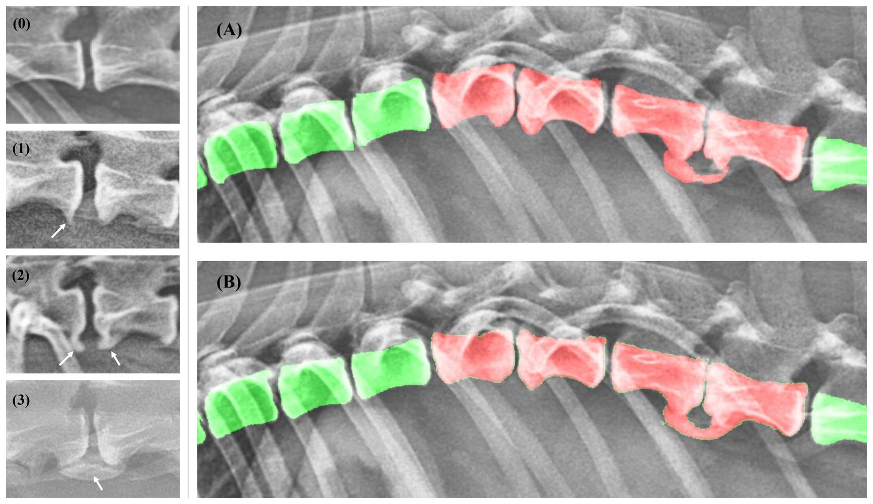
Spondylosis deformans grading (grades 0–3, arrows = bony spurs) and a modified attention U-Net → manual (A) vs. automated (B) segmentation of vertebral bodies (green) and spondylosis (red) on canine lateral X-rays · Frontiers in Veterinary Science, 2024 (JCR top 13%)
Spondylosis deformans grading (grades 0–3, arrows = bony spurs) and a modified attention U-Net → manual (A) vs. automated (B) segmentation of vertebral bodies (green) and spondylosis (red) on canine lateral X-rays · Frontiers in Veterinary Science, 2024 (JCR top 13%) Paper ↗
1 / 3
02

Physics-Informed Neural Networks for Ultrasound Imaging

Physics-guided reconstruction, inverse problems, quantitative imaging

Physics-informed neural networks that embed wave propagation, beamforming, and image-formation models into self-/unsupervised learning for high-fidelity ultrasound reconstruction, physics-guided inverse modeling for artifact reduction, and learning-based quantitative and perfusion ultrasound.

Physics-informed learningUnsupervised beamformingInverse problemsQuantitative imaging
Unsupervised deep coherence learning: a network trained to predict signals coherent across steering angles → clutter-suppressed single plane-wave carotid images (DAS vs. DL-DCL) · Ultrasonics, 2024 (JCR top 11%)
Unsupervised deep coherence learning: a network trained to predict signals coherent across steering angles → clutter-suppressed single plane-wave carotid images (DAS vs. DL-DCL) · Ultrasonics, 2024 (JCR top 11%) Paper ↗
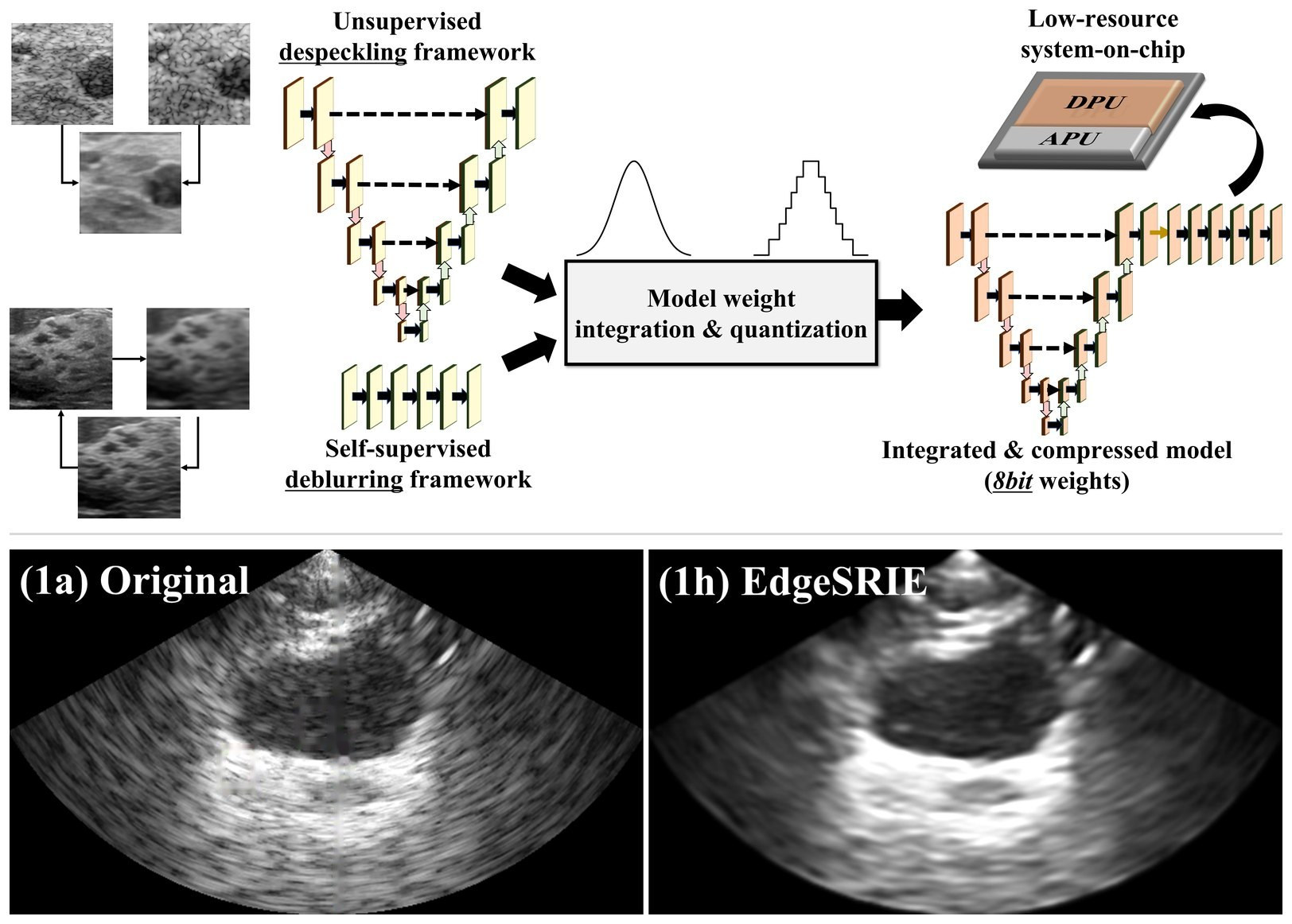
Unsupervised despeckling and self-supervised deblurring fused into one 8-bit model for a low-resource SoC → real-time speckle-reduced bladder imaging on a portable device · Medical Physics, 2026
Unsupervised despeckling and self-supervised deblurring fused into one 8-bit model for a low-resource SoC → real-time speckle-reduced bladder imaging on a portable device · Medical Physics, 2026 Paper ↗
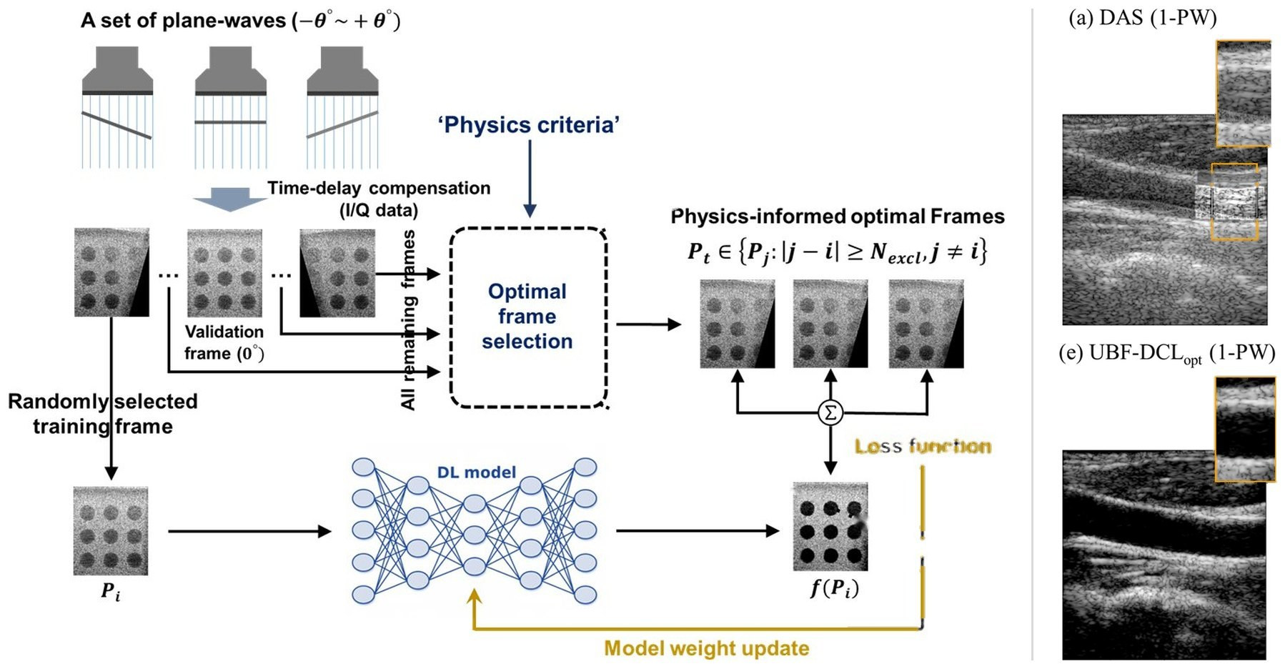
Physics-informed optimal-frame selection for an unsupervised coherence loss → clutter-suppressed single plane-wave carotid imaging (DAS vs. UBF-DCLopt) · Diagnostics, 2025 (JCR top 17%)
Physics-informed optimal-frame selection for an unsupervised coherence loss → clutter-suppressed single plane-wave carotid imaging (DAS vs. UBF-DCLopt) · Diagnostics, 2025 (JCR top 17%) Paper ↗
1 / 3
03

Physical AI for Wearable & Robotic Ultrasound

Active perception, sim-to-real, edge AI

Closed-loop robotic ultrasound guidance with active scanning policies, self-supervised adaptation across operators, devices, and patients, and data-/compute-efficient learning with hardware-aware design for edge devices.

Robotic ultrasoundWearable imagingSim-to-real transferEdge AI & SoC
AI-guided beamforming for echocardiography: an echo foundation model maps frames into a latent space where the probe angle is steered toward the standard view (unoptimized → standard) · ongoing work with SMART Centre, MIT
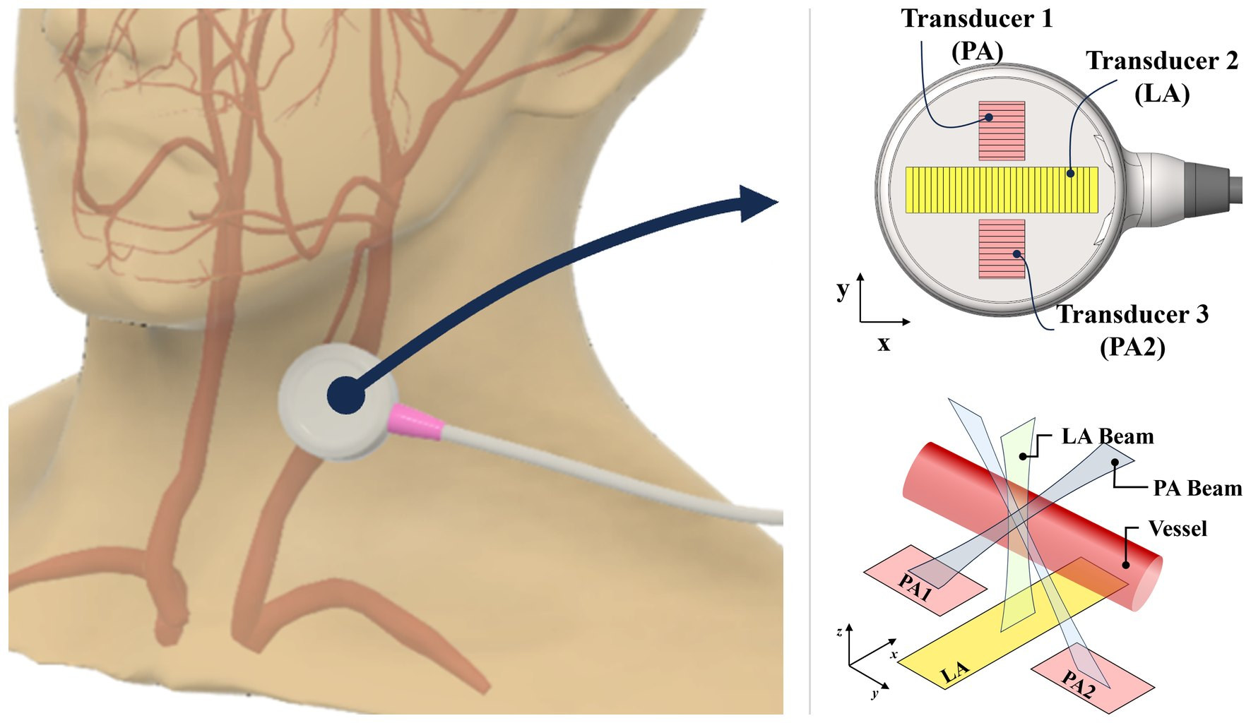
Wearable cross-shaped ultrasound array worn on the neck → a linear array plus two orthogonal phased arrays whose crossed beams enable angle-independent vector Doppler flow monitoring · IEEE TIM, 2026 (JCR top 5.6%)
Wearable cross-shaped ultrasound array worn on the neck → a linear array plus two orthogonal phased arrays whose crossed beams enable angle-independent vector Doppler flow monitoring · IEEE TIM, 2026 (JCR top 5.6%) Paper ↗
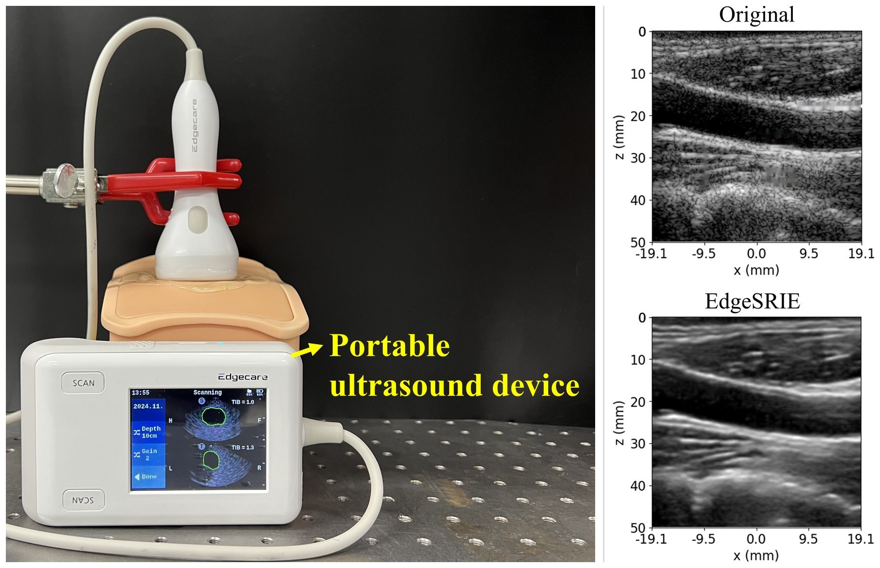
EdgeFlow UH-10 portable ultrasound scanner → Original vs. EdgeSRIE on an in vivo carotid scan (PICMUS); the deployed 8-bit model runs at 64 frames/s on the device-class low-resource SoC · Medical Physics, 2026
EdgeFlow UH-10 portable ultrasound scanner → Original vs. EdgeSRIE on an in vivo carotid scan (PICMUS); the deployed 8-bit model runs at 64 frames/s on the device-class low-resource SoC · Medical Physics, 2026 Paper ↗
1 / 3